Home
/ Diagram Of Hip.and Back.muscles : Hip Strains Orthoinfo Aaos, The image below shows the bones from the back side of the hand.
Diagram Of Hip.and Back.muscles : Hip Strains Orthoinfo Aaos, The image below shows the bones from the back side of the hand.
Diagram Of Hip.and Back.muscles : Hip Strains Orthoinfo Aaos, The image below shows the bones from the back side of the hand.. Muscles of the upper limb (deltoid, biceps, forearms). It is opposite from the chest, and the vertebral column runs down. Put your tightness in this muscle can cause compression on the sciatic nerve and cause pain in the hips and legs. The hip muscle diagram below shows a number of the muscles we will be discussing in the next sections. As a result, you build a more muscular back and can help prevent back pain.
Hip muscles and tendons march 19 2019 by luqman. Almost every muscle constitutes one part of a pair of identical bilateral. Other muscles are small and cover much less space. Deadlift muscles will include knee, hip, and back extensors, which primarily include the quads, glutes, and spinal erectors. Muscles of the hip and lower limb.
Tight Hip Flexors And Back Pain Knee And Foot Pain The Biomechanics Method from www.thebiomechanicsmethod.com Lower back muscles below the shoulder blade. Muscles of the hip and lower limb. Hip muscles and tendons march 19 2019 by luqman. Muscles of the hip & thigh (quadriceps, hips). This article looks at the anatomy of the back, including bones, muscles, and nerves. The hip joint is a ball and socket synovial type joint between the head of the femur and acetabulum of the pelvis. Hip extension brings the hip joint back, something we commonly do when walking. Now that you watched the video, you.
This article looks at the anatomy of the back, including bones, muscles, and nerves.
Muscles in the human body (pectoralis major, abdominals, obliques). Human muscle system, the muscles of the human body that work the skeletal system, that are under voluntary control, and that are concerned with movement, posture, and balance. Key facts about hip muscles. Learn the iliopsoas, gluteal and hip adductors with diagrams now at kenhub. Diagram of muscles and anatomy charts. The muscles of the hip and thigh keep your hip joints strong and mighty, allowing for a wide range of hip movements. Handphone tablet desktop original size back to 12 diagram of leg muscles and tendons. Lower back muscles below the shoulder blade. Sit on the floor with your legs extended straight in front of you 2. This is a table of skeletal muscles of the human anatomy. The back's muscles start at the top of the back (named the cervical vertebrae) and go to the tailbone (also named the coccyx). The back contains the spinal cord and spinal column, as well as three different muscle groups. Now that you watched the video, you.
Now that you watched the video, you. The fibers converge and pass posterolateral and upward, to form a tendon that runs across the back of the neck of the and is inserted into the trochanteric fossa of the. Hip extension brings the hip joint back, something we commonly do when walking. Common hip and back pain causes include injury to muscles from overuse disc injurydegeneration or spinal stenosis. Back muscles anatomy lower back muscles anatomy human anatomy.
Today S Focus Is Hip And Ankle Joint Muscles And Actions I Will Remember The Following Actions Of The Hip Leg Muscles Anatomy Hip Muscles Hip Muscles Anatomy from i.pinimg.com The back contains the spinal cord and spinal column, as well as three different muscle groups. The image below shows the bones from the back side of the hand. Muscles of the upper limb (deltoid, biceps, forearms). Back muscles anatomy lower back muscles anatomy human anatomy. Bend your right leg 3. Many conditions and injuries can affect the back. Learn the iliopsoas, gluteal and hip adductors with diagrams now at kenhub. There are anterior muscles diagrams and posterior muscles diagrams.
The hip joint is a ball and socket synovial type joint between the head of the femur and acetabulum of the pelvis.
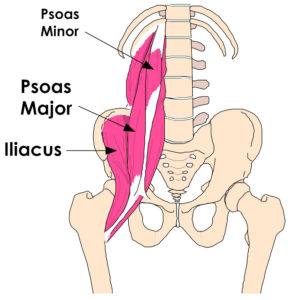
These muscles form the pelvic diaphragm which supports and maintains the position of the pelvic ilium, sacrum, coccyx and lumbodorsal fascia. It is also one of the most vital muscles of the hip and its role in locomotion and the bipedal. Iliacus, psoas major, and psoas minor main function: Because this muscle inserts onto the back of the greater trochanter, it produces lateral rotation at the hip. Anatomy back anatomy bones gross anatomy human body anatomy muscle anatomy lower back muscles anatomy shoulder anatomy muscle diagram anatomy images. There are anterior muscles diagrams and posterior muscles diagrams. The levator ani muscle along with a second muscle forms the pelvic floor. The red lines show where the tendons attach the muscles to the bones. Muscles of buttock, hip and pelvis laminated anatomy chart. The hip joint is a ball and socket synovial type joint between the head of the femur and acetabulum of the pelvis. The back's muscles start at the top of the back (named the cervical vertebrae) and go to the tailbone (also named the coccyx). The image below shows the bones from the back side of the hand. This is a table of skeletal muscles of the human anatomy.
Hip muscles and tendons march 19 2019 by luqman. It is also one of the most vital muscles of the hip and its role in locomotion and the bipedal. Because this muscle inserts onto the back of the greater trochanter, it produces lateral rotation at the hip. Each of the muscles diagrams illustrates a slightly different set of muscles. The red lines show where the tendons attach the muscles to the bones.
Piriformis Syndrome from www.spineuniverse.com The red lines show where the tendons attach the muscles to the bones. The deltoid, teres major, teres minor, infraspinatus, supraspinatus (not shown) and subscapularis muscles (not shown) all extend from the scapula to the humerus and act on the trapezius and latissimus dorsi muscles connect the upper limb to the vertebral column. These muscles form the pelvic diaphragm which supports and maintains the position of the pelvic ilium, sacrum, coccyx and lumbodorsal fascia. Key facts about hip muscles. Muscles of the hip & thigh (quadriceps, hips). Other muscles are small and cover much less space. Back muscles anatomy lower back muscles anatomy human anatomy. It joins the lower limb to the pelvic girdle.
Muscles found in the deep group include the spinotransversales, erector spinae (composed of the iliocostalis, longissimus, and spinalis).
Muscles of buttock, hip and pelvis laminated anatomy chart. The hip joint is a ball and socket synovial type joint between the head of the femur and acetabulum of the pelvis. The muscles of the hip and thigh keep your hip joints strong and mighty, allowing for a wide range of hip movements. Iliacus, psoas major, and psoas minor main function: The levator ani muscle along with a second muscle forms the pelvic floor. Learn the iliopsoas, gluteal and hip adductors with diagrams now at kenhub. The image below shows the bones from the back side of the hand. The hip muscle diagram below shows a number of the muscles we will be discussing in the next sections. You can protect the back muscles by bending from the hip and. Back pain is the most common type of chronic is it any wonder that many consider the deadlift as the king of all exercises? Common hip and back pain causes include injury to muscles from overuse disc injurydegeneration or spinal stenosis. Back muscles are divided into two specific groups: It joins the lower limb to the pelvic girdle.

